
A BIOS REU, Times Two
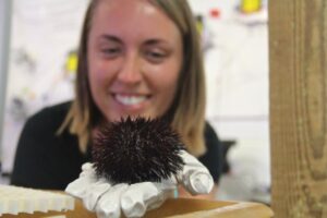
Chloe Emerson initially came to BIOS in the fall of 2014 for the Research Experiences for Undergraduates (REU) internship program funded by the National Science Foundation. As a Wellesley College senior working to complete her major in Biology and minor in Philosophy, Emerson already found developmental biology and stem cell research fascinating. At BIOS, these interests crystalized as she began to study sea urchins in Andrea Bodnar’s Molecular Discovery Laboratory, leading her down a path in regenerative biology that she hardly could have imagined two years ago.
A Toolkit to Measure Regeneration
Emerson spent her initial twelve-week REU internship in 2014 working with Bodnar and BIOS postdoctoral scholar Helena Reinardy to optimize a method measuring regeneration of tube feet and spines in sea urchins, and then using the method to examine a biochemical pathway involved in regeneration.
Sea urchins, sea stars and sea cucumbers all belong to a group of invertebrates remarkably capable of regenerating tissues such as nerve and muscle, and even their calcium-carbonate skeletons. Measuring the regeneration of sea urchin spines and fleshy tube feet provides insight into the biological underpinnings of two potentially different processes, and understanding how animals regrow body parts could one day open new possibilities in regenerative therapies for humans.
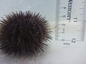
A sea urchin’s nearly translucent, sticky tube feet (pictured extended, above) can reach beyond the spines to sense the surrounding environment and attach to underwater surfaces, but are completely retracted when the sea urchin is removed from the water.
While sea urchin spines can be measured by hand using calipers, sea urchins quickly retract their tube feet when removed from the water or under threat. Therefore underwater photography and a protocol for image analysis were required to measure length of tube feet as they regrew. Amputating a whole row of tube feet to measure regrowth was no easy task, as the sea urchins retract all their tube feet each time a single one was clipped, making for some long days at the beginning of each experiment. However, once the team could reliably track and compare regrowth of clipped spines and tube feet, Emerson could use the method to investigate the biochemistry behind regeneration.
An interesting initial testing ground was the Notch signaling pathway, a cell-to-cell signaling system present in nearly all animals, which is often involved in development and regeneration. The sea urchin genome has the genes for Notch signaling, but the role of these genes in adult sea urchin regeneration had never been tested. After clipping the spines and tube feet, a subset of sea urchins were injected with a chemical inhibitor known to block the action of the Notch signaling pathway. The inhibitor stunted regeneration of both tube feet and spines in the urchins, and turned off the genes involved in this cell-to-cell signaling pathway. Together, the physical and genetic data showed that Notch signaling in sea urchins plays an essential role for regrowth of both skeleton and tissue. The results were published in the scientific journal PLOS One in August 2015.
In recognition of her accomplishments during the twelve-week REU program, Emerson received BIOS’s inaugural REU Supplemental Award to support a second summer of research addressing related research questions.
With Rising Carbon Dioxide Emissions, Does Spine Regeneration Suffer?
Reuniting in 2015, Emerson, Reinardy and Bodnar wanted to apply their method for measuring spine regrowth to better understand how shifting conditions in the environment might impact the regenerative capabilities of sea urchins.
The twenty-seven year Bermuda Atlantic Time-series Study (BATS) has clearly documented how carbon dioxide absorbed by the ocean has lowered seawater pH over the last three decades. With the current pace of global carbon dioxide emissions, ocean pH will continue to drop in the coming years. “From the beginning, something the three of us had talked about was that ocean acidification is probably really relevant to these organisms,” said Emerson. That’s because sea urchin skeletons and spines are composed of a form of calcium carbonate that is particularly sensitive to the changing ocean chemistry.
At BIOS, Bodnar’s molecular biology lab is in the unique position of being surrounded by colleagues who are monitoring acidification in the ocean, have a deep understanding of the chemistry involved, and also have experience manipulating it experimentally in the lab. As such they were lucky to have good advice and practical help as they set up tanks bubbled with carbon dioxide to create “moderate” and “high” acidification conditions for the sea urchins (pH of 7.7 and 7.5, respectively, compared to Ferry Reach water’s natural pH of 8.1). Coral biologist Samantha dePutron lent instruments to control the flow of gasses, and zooplankton ecologist Amy Mass helped get the seawater systems up and running. BIOS scientist Nick Bates advised on the water chemistry, and his lab analyzed weekly water samples from all three tanks to provide essential data to monitor and accurately describe the experimental conditions.
Over two months, the sea urchins in all three seawater conditions (high acidification, moderate acidification, and ambient seawater) regrew their spines at equivalent rates. However, when Emerson tested how the sea urchins were expressing genes involved in biomineralization – the biochemical pathways involved in concentrating calcium and carbonate compounds from the water to build new spines – there was different gene expression in the sea urchins kept in acidified water, suggesting more of a struggle at the biochemical level.
“We were pretty amazed that under these stressful acidified conditions they were able to keep regrowing their spines,” said Emerson, “but when we were handling the urchins, it seemed the spines would sort of crumble in our hands.” Perhaps despite the sea urchin’s best effort to regrow spines, the ocean acidification conditions were compromising spine integrity?
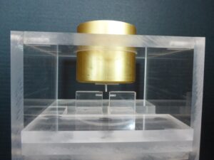
Bruce Williams’ invention, pictured here with a sea urchin spine balanced between the platforms, ready for the “spine snap test”.
Necessity, the Mother of Invention
To test whether spines really did break more easily when animals were kept in acidified seawater, a formal spine strength test had to be developed. BIOS Laboratory Operations Technician Bruce Williams was called in to help build an instrument that could determine the breaking point of individual spines.
Bruce built an elegant contraption that came to be known informally as “The Urchinator.” It is designed so that a single spine lies across a gap between two platforms, and a container with a bar on the bottom is delicately balanced on the spine’s midpoint. The scientists would slowly drop tiny lead balls into the container to determine the amount of weight an individual spine could bear before breaking.
“We were carefully balancing hundreds of spines, just wondering if there actually is a real difference, and could this crude method even capture it?” Emerson remembered. At this point Reinardy’s BIOS postdoctoral position had ended and she was communicating with Emerson from a new job in Svalbard, Norway. However, Reinardy’s belief in the project never waivered and she cheered on the tedious measurements from afar.
Animals from each of the three experimental tanks had ten regrown spines and ten previously existing spines subjected to the test, so Emerson ended up delicately balancing and testing the breaking point of over 360 spines. When she examined the results, she found that spines from sea urchins living in acidified seawater broke under significantly less weight – and the more acidic the seawater, the more fragile the spine.
Taken together, Emerson thinks these results suggest that sea urchins must heavily prioritize regrowing their spines to protect their body and internal organs from predators, even if chemical conditions are subpar and gene expression shows the regrowth is a costly endeavor for sea urchins.
A New Perspective on Science
The challenges and new ideas that arose over the course of two research internships inspired Emerson, and she continues to immerse herself in the field of regenerative biology. Last year, she spent three weeks studying comparative regenerative biology at the Mount Desert Island Biological Laboratory in Bar Harbor, ME, and attended the Comparative Biology of Tissue Repair, Regeneration and Aging Symposium that was hosted there. Emerson also submitted an abstract describing her REU results to the Molecular and Cellular Basis of Growth and Regeneration Keystone Symposium. Not only was the abstract accepted, but also she was invited to give a talk – a rare honor for an undergraduate student. With travel support from her REU Supplemental Award, Emerson presented her work at the Symposium in Breckenridge, CO in January 2016.
“This whole experience has been extremely valuable to me, in large part because Andrea and Helena were amazing mentors,” said Emerson. “Whenever I had a question they were there to talk me through the options, but they also gave me space to do the work and think on my own. They read every draft I wrote and offered a million comments, which really helped me develop as a writer. And because the second research experience involved so much troubleshooting, that exposed me even more to what science is really like!”
While there’s no telling exactly where her research interests will take her next as she investigates graduate programs, the work done in her double REU has already provided Emerson entree into the field of regenerative biology, and it looks like she intends to stay there.