
Medical Imaging and Marine Bacteria
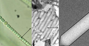
The smallest photosynthesizing organisms in the ocean, cyanobacteria, have the potential to transform the field of medical imaging and are a topic of interest for BIOS molecular biologist Julius Christopher Barsi. Cyanobacteria (shown here at left under a light microscope) contain a unique feature within their cells: microscopic nanostructures called gas vesicles (shown in the middle under low resolution electron micrograph and at right under high resolution electron micrograph). Barsi believes these hollow structures can be harnessed for non-invasive imaging, such as MRIs, and present a safer alternative to the contrast agents that are currently being used. Photo credit: Herzfield/Melanson/DeRosier
In the U.S. alone, approximately 40 million people receive Magnetic Resonance Imaging (MRI) exams each year. These procedures use computer-generated radio waves and strong magnets to create detailed 3-D pictures of areas inside the human body, helping doctors diagnose a variety of conditions, such as blood clots, bone infections, or compressed discs in the spine.
In about a third of cases, MRIs involve the use of a “contrast agent” to improve the clarity of the images of your body’s internal structures, particularly in areas with abnormally high levels of blood supply, such as in tumors.
“The contrast agent requires the element gadolinium, which is retained in the bodies of chemotherapy patients, causing further health damage,” said BIOS molecular biologist Julius Christopher Barsi. On one hand there is a pressing need to accurately visualize tumors, on the other hand the only way to do that is harmful for cancer patients. “By combining a recent discovery in the ocean with what we learned during the pandemic about using nanoparticles as delivery agents for mRNA, such as with COVID-19 vaccines, the stage is set to increase the safety of tumor visualization.”
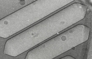
By combining the discovery of these gas vesicles with “what we learned during the pandemic about using nanoparticles as delivery agents for mRNA, such as with COVID-19 vaccines,” Barsi feels “the stage is set to increase the safety of tumor visualization” in cancer patients. The goal would be to utilize the gas vesicles to develop a “therapeutic acoustic reporter gene,” leveraging recent advances in nanoparticle delivery technology to target a specific “address” in the body (i.e., the tumor). Afterward, a sonogram would collapse the gas vesicles, removing them entirely. Photo credit: Herzfield Group.
Visualizing with Gas Vesicles
Cyanobacteria, commonly known as blue-green algae, are the smallest photosynthesizing organisms in the ocean. Found in all environments, they are particularly common in the open ocean, where some cyanobacteria play important roles as “nitrogen fixers,” converting nitrogen gas into other more biologically useful compounds.
One of the properties that makes cyanobacteria unique, and is poised to transform the field of medical imaging, are microscopic organelles within the cell called gas vesicles. These hollow protein nanostructures trap gas to improve buoyancy and help the bacteria position themselves within the water column. Importantly, they also possess unique physical properties that can be harnessed for non-invasive imaging.
“When you have an x-ray taken of your lungs, there is a clear image because of the contrast between surrounding tissue and the air within your lungs,” Barsi said. In the same manner, the minute amount of gas trapped by these microscopic gas vesicles within the cell improves tissue visualization in ultrasounds, and it can also enhance the capabilities of MRIs.
Combining the concept described above with an mRNA delivery system to humans results in what molecular biologists refer to as “therapeutic acoustic reporter genes,” or engineered clusters of genes that can be used for therapeutic medical imaging. When you administer an mRNA vaccine, the cells of the body generate the antigen. Similarly, the mRNA of a gas vesicle could be administered just like current vaccines, encoded with a small protein that can be directly visualized with ultrasound and serve as a safe alternative to existing MRI contrast agents.
Barsi feels that medical applications of gas vesicles from marine cyanobacteria are underexplored. He also notes there have been recent advances outside of marine genomics that make this an ideal topic for further investigation. Contemporary vaccines employ liposome nanoparticles as a delivery agent, necessary to “protect the cargo” of the vaccine.
The ubiquitous testing and usage of nanoparticles as a delivery agent during the past few years has demonstrated their efficacy and led to enhanced capabilities, such as targeting a specific “address” within the body. This has potential medical applications in the field of cancer treatments. For example, across a large number of tumors, the folate receptor is overexpressed. Folate receptors bind to folic acid like a “lock-and-key” mechanism. By adding folic acid to the delivery agent, we can facilitate its passage into the cell and ensure that gas vesicle mRNA ends up where it is most needed for imaging (i.e., in the tumors themselves).
The best part Barsi saves for last. A person who has been injected with an acoustic reporter gene for the purpose of imaging a tumor, would be reassured to know that a quick sonogram of the same area will collapse the vesicles, removing them entirely.
A Survey of the Sargasso Sea
Barsi’s goal is to attract funding and collaborators that will allow him to conduct a survey of organisms in the Sargasso Sea, with the idea of discovering gas vesicles with novel physical properties or variations of known gas vesicles.
Simply sequencing DNA found in ocean water would be like searching for a needle in a haystack. This is why Barsi has designed an assay to specifically discover gas vesicle gene clusters across phyla.
Whereas others are attempting to improve the acoustic properties and expression characteristics of known gas vesicles through synthetic manipulation, Barsi said: “I want to explore what is already present in nature and see what evolution has given us. Even if you aren’t going to use the whole DNA sequence, you might encounter a small part, or an allele that is particularly valuable. But we need to start with a survey of what’s out there.”